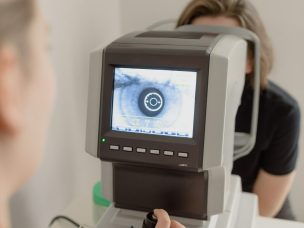
Optical coherence tomography angiography (OCT-A) is a new, non-invasive imaging technique used to evaluate retinal and choroidal microvasculature. This cross-sectional study, published in the Annals of Medicine and Surgery, sought to determine OCT-A’s diagnostic accuracy in assessing the morphological features and clinical activity of macular neovascularization (MNV) in wet age-related macular degeneration (AMD).
The researchers’ study lasted for 15 months, during which macular OCT-A images were gathered from 55 patients with wet AMD using a swept-source OCT-A device. These images were then used to analyze morphological characteristics and other metrics. An extensive qualitative description of detected MNV was noted, along with the area of the lesion in square millimeters.
Ultimately, it was found that the modality’s sensitivity was 85% for type 1 MNV lesions; 100% for type 2, mixed type 1 and 2, and type 3 MNV lesions; and 92% for unclassified fibrotic lesions. Moreover, its sensitivity was 84.6% for active lesions and 96.8% for inactive lesions. The researchers concluded that OCT-A is a safe, accurate, and reproducible technique for diagnosing and monitoring wet AMD [1].
Source:
[1] Ahmed, M., Syrine, B. M., Nadia, B. A., Anis, M., Karim, Z., Mohamed, G., Hachemi, M., Fethi, K., & Leila, K. (2021). Optical coherence tomography angiography features of macular neovascularization in wet age-related macular degeneration: a cross-sectional study. Annals of Medicine and Surgery, 70, 102826. https://doi.org/10.1016/j.amsu.2021.102826


