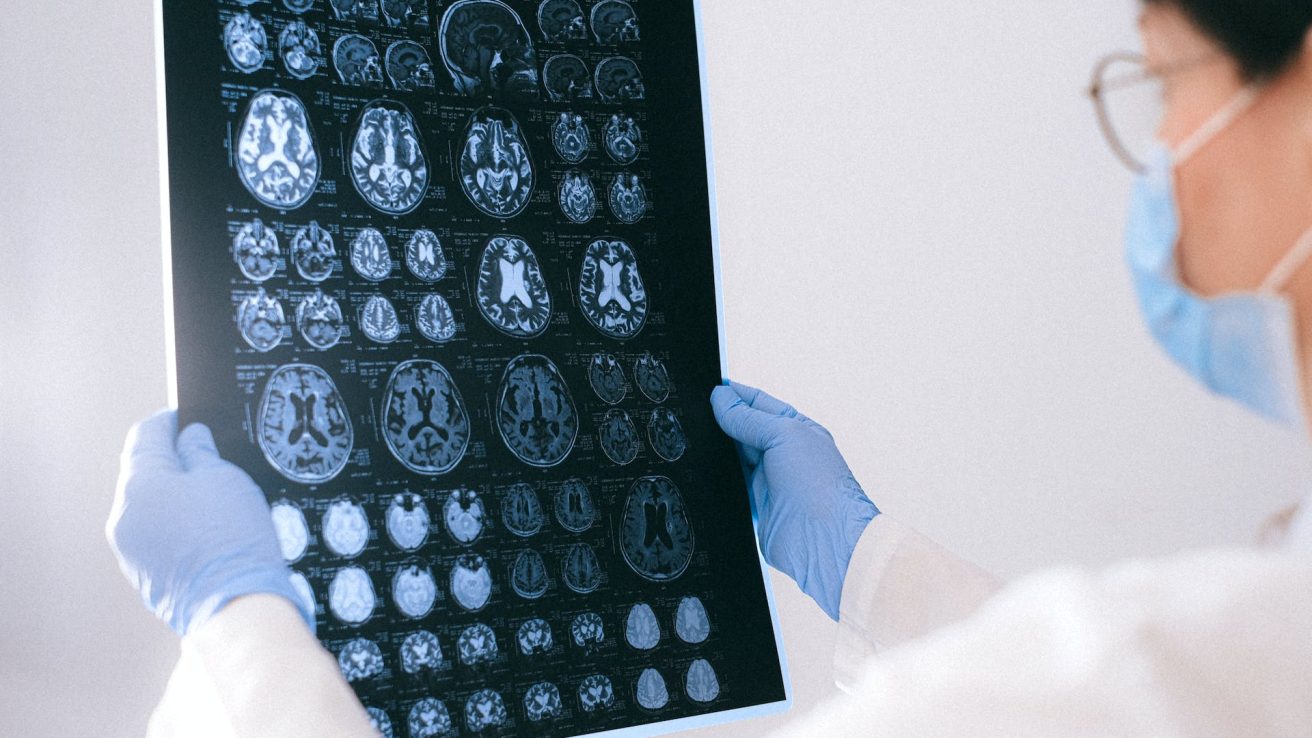
Brain MRI volumetric measurements are a better predictor of cognitive performance than neurofilament light chain levels in multiple sclerosis, according to a recent 10-year follow-up study.
Cognitive impairment is an important factor in disability due to multiple sclerosis (MS). The neurofilament light chain (NfL) is a promising biomarker for neuroaxonal damage. Research has correlated increased NfL levels with brain and spinal cord volume loss. Several studies have linked magnetic resonance imaging (MRI) variables to cognitive impairment; however, varying results have been observed regarding the association between NfL and cognitive decline.
A study in Acta Neurologica Scandinavica assessed the association between NfL and cognitive function over 10 years in comparison to MRI and clinical data in MS patients.
Study Population
A total of 41 MS patients were enrolled in this prospective longitudinal cohort study. The mean age was 41.6 years, and 73% were female. The median disease duration was 60 months at the time of diagnosis, with 83% being relapsing–remitting MS cases and 17% being secondary progressive MS cases.
Significant Associations Found Between NfL Levels and Symbol Digit Modalities Test
The symbol digit modalities test (SDMT) was used to assess cognitive processing speed. At baseline, a significant cross-sectional correlation (p=0.005) was observed between SDMT and log-transformed serum NfL (sNfL). Meanwhile, a slightly lower correlation (p=0.030) was observed between SDMT and log-transformed cerebrospinal fluid NfL (cNfL). Significant cross-sectional correlations were also observed between log-transformed baseline sNfL and SDMT at 5-year (p=0.015) and 10-year (p=0.009) time points and between log-transformed baseline cNfL and SDMT at the 10-year point (p=0.023), but not at five years.
Longitudinal analyses revealed a significant association between baseline sNfL and SDMT during the 10-year period (p<0.001). The expected baseline SDMT decreased by 9.5 points for each one-unit higher log sNfL (p=0.002), but the association with longitudinal change in SDMT was not statistically significant from baseline to 5 years or from baseline to 10 years.
Significant Associations Revealed Between MRI Volumes and SDMT
At baseline, significant cross-sectional correlations were observed for T1 and T2 lesion volumes (p<0.001), whole brain volume (p=0.002), and gray matter volume (p=0.002) with SDMT. The baseline T1 lesion volume (p=0.001 and p<0.001), T2 lesion volume (p=0.004 and p=0.001), gray matter volume (p<0.001 and p=0.002), and whole brain volume (p<0.001 and p=0.002) all correlated significantly with SDMT at 5-year and 10-year points.
Longitudinal analyses revealed a significant association between T1 lesion volume and baseline SDMT. The expected baseline SDMT decreased by 5.1 points for each one-unit higher square root T1 lesion volume (p<0.001), with an increased rate of decline in SDMT from baseline to 10 years (p=0.002).
Associations Found Between Expanded Disability Status Scale and SDMT at Baseline and Five Years
The baseline Expanded Disability Status Scale (EDSS) significantly correlated with the baseline SDMT (p=0.005) and 5-year SDMT (p<0.001), but not with 10-year SDMT. Longitudinal analysis showed a significant correlation between EDSS and SDMT (p<0.001) and between baseline EDSS and a decrease in SDMT from baseline to 5 years (p<0.001).
MRI Volume Metrics Outperform NfL Levels in Predicting Cognitive Decline in MS
According to the model fit statistics analysis, all baseline MRI volumes (whole brain, gray matter, and T1 and T2 lesion volumes) and EDSS demonstrated stronger associations with longitudinal SDMT than those shown by baseline cNfL and sNfL. Baseline T1 lesion volume showed the strongest association with SDMT. Therefore, although NfL levels were associated with cognitive functioning in MS, MRI volumes additionally correlated with and predicted the rate of cognitive decline better than NfL.
Source:
Bhan, A., Jacobsen, C., Dalen, I., Alves, G., Bergsland, N., Myhr, K., Zetterberg, H., Zivadinov, R., & Farbu, E. (2023). Neurofilament and Brain Atrophy and Their Association with Cognition in Multiple Sclerosis: A 10-Year Follow-Up Study. Acta Neurologica Scandinavica, 2023, 1–10. https://doi.org/10.1155/2023/7136599


