The emergence of macular IGH-like lesions is a reversible and rare adverse effect following photochemotherapy in vitiligo patients.
Phototherapy is a safe and effective treatment for stable vitiligo in the pediatric population. A rare side effect of phototherapy is the formation of small and well-defined depigmented macules, specifically in skin regions with exposure to ultraviolet radiation. These macules are termed idiopathic guttate hypomelanosis (IGH)-like lesions, leucoderma punctatum, and confetti-like hypopigmentation. The scientific literature regarding these macules is limited to case reports and case series.
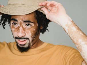
This study, published in the Journal of Dermatology, Venereology, and Leprology, recruited five children diagnosed with stable vitiligo. The children received photochemotherapy comprising topical psoralen followed by sun exposure (PUVAsol) for a variable duration. The investigators observed multiple, small, and well-defined depigmented macules of 1–5 mm size within and in the surrounding regions of vitiliginous skin. The macules were restricted to skin areas with exposure to photochemotherapy. The development of macules was observed after a few months of starting the PUVAsol therapy. The quantity of macules increased with the continuation of photochemotherapy. However, the investigator observed the re-pigmentation of vitiligo lesions of a mild to moderate degree.
In contrast to larger and chalky white lesions of vitiligo, the depigmented macules are smaller and have a brighter white hue. While studying the macules using dermoscopy, the investigator observed a cloudy appearance, a lack of a pigmentary network, and the presence of numerous convex outpouchings. The biopsy findings of these macules from a single patient include hyperkeratosis, mild papillomatosis, the near absence of pigmentation in the basal layer, and the flattening of the rete ridges of the macules.
A prominent and abrupt transition was observed between non-pigmented and pigmented basal layers. The study findings also suggested a proportional increase in the density of macules upon exposure to the increasing density of phototherapy. Based on the characteristic features, the authors suggest the lesions to be IGH instead of being IGH-like macules, owing to similar histological findings. The appearance of the lesions is not limited to children and psoralen-induced photochemotherapy. During the follow-up period of 1–3 months, there were no changes in the extent of re-pigmentation, vitiligo patches, or IGH-like macules.
In summary, healthcare providers should be aware of lesions and resultant dyschromia in phototherapy. Phototherapy should be discontinued in cases of worsening vitiligo lesions. This study warrants the further investigation of lesions, a longer duration follow-up period, and a thorough histopathological investigation of lesions in all study subjects.
References
Mehta, N., Yadav, D., & Bhari, N. (2022). Idiopathic guttate hypomelanosis-like macules co-localizing to sites of topical photochemotherapy for vitiligo in children: Clinical and dermoscopic findings. Indian Journal of Dermatology, Venereology and Leprology, 1-3. https://doi.org/10.25259/ijdvl_273_2022