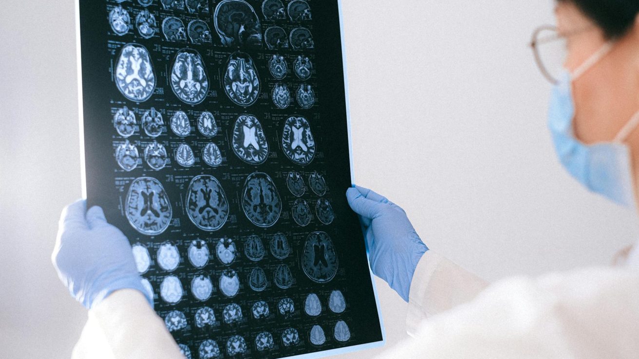
A prospective study shows that it is possible to differentiate between relapsing–remitting multiple sclerosis and aquaporin-4 antibody-positive neuromyelitis optica spectrum disorder with high accuracy using MRI imaging. However, differentiating between neuromyelitis optica spectrum disorder and myelin oligodendrocyte glycoprotein antibody-associated disease using imaging methods is relatively challenging.
There is a lot of overlap between clinical and imaging manifestations of neuromyelitis optica spectrum disorders (NMOSD), multiple sclerosis (MS)/ relapsing–remitting MS (RRMS), and myelin oligodendrocyte glycoprotein antibody-associated disease (MOGAD). Serological testing for aquaporin-4 (AQP4) antibody (Ab) and MOG-Ab is a highly specific method for differentiating between the conditions. However, there are some challenges, including variable sensitivity and the unavailability of these tests in many cases. The MOG-Ab test produces false-positive results in 28% of cases when used indiscriminately. Hence, challenges remain in differentiating NMOSD from RRMS and MOGAD.
Magnetic resonance imaging (MRI) may provide valuable insights in many instances, helping to differentiate between the conditions. This study, published in the journal Neurology, focuses on differentiating between the conditions using MRI.
MRI Imaging Shows Significant Differences Between the Conditions
The study enrolled 91 patients, including 30 APQ4-NMOSD, 31 RRMS, and 30 MOGAD, along with 34 healthy subjects. For differentiating between RRMS and NMOSD, the most reliable observations were lesions with the central vein sign (CVS) (84% vs. 33%, respectively), with an accuracy of 91%, specificity of 88%, and sensitivity of 93%, p < 0.001.
The second-most accurate distinquisher between RRMS and NMOSD was the number of cortical lesions, with accuracy, specificity, and sensitivity of 84%, 90%, and 77%, respectively, p = 0.002. This was followed by white matter lesions, with accuracy, specificity, and sensitivity of 78%, 84%, and 73%, respectively, p = 0.001. Combining CVS, cortical lesions, and the optic nerve magnetization transfer ratio could help reach an accuracy, specificity, and sensitivity of 95%, 92%, and 97%, respectively, p < 0.001.
Further, the study found that when differentiating RRMS from MOGAD, white matter lesions had an accuracy, specificity, and sensitivity of 94%, 94%, and 93%, respectively, p = 0.006. High cortical lesion numbers combined with higher retinal nerve fiber layer thickness could differentiate between RRMS and MOGAD with accuracy, specificity, and sensitivity of 84%, 92%, and 77%, respectively, p < 0.001.
The Bottom Line
This study demonstrated the possibility of differentiating between these conditions with high accuracy using MRI imaging. It was found that brain cortical and white matter lesions could help differentiate RRMS from antibody-mediated conditions such as NMOSD and MOGAD. The magnetization transfer ratio of the optic nerve was another reliable way of differentiating RRMS from NMOSD. However, the study also found that differentiating between AQP4-NMOSD and MOGAD is relatively challenging due to their many similarities. Nonetheless, the presence of at least one cervical cord lesion may help.
This study could help healthcare professionals differentially diagnose these conditions when antibody testing is not available or its findings are questionable. However, this study has one significant limitation: it was done in nonacute patients with established diseases. It is unclear if these imaging parameters can help distinguish between the conditions at onset. Additionally, it remains unclear if these differences in MRI are also seen with changes over time.
Source:
Cortese, R., Carrasco, F. P., Tur, C., Bianchi, A., Brownlee, W., De Angelis, F., De La Paz, I., Grussu, F., Haider, L., Jacob, A., Kanber, B., Magnollay, L., Nicholas, R., Trip, A., Yiannakas, M., Toosy, A., Hacohen, Y., Barkhof, F., & Ciccarelli, O. (2023). Differentiating multiple sclerosis from AQP4-Neuromyelitis optica spectrum Disorder and MOG-Antibody Disease with imaging. Neurology, 100(3). https://doi.org/10.1212/wnl.0000000000201465


